A mammogram is an x-ray exam of the breast that’s used to detect and evaluate breast changes. Today’s x-ray machines used for mammograms expose the breast to much less radiation compared with those used in the past.
Screening mammograms are x-ray exams of the breasts that are used for women who have no breast symptoms or signs of breast cancer. The goal of a screening mammogram is to find breast cancer when it’s too small to be felt by a woman or her doctor, which gives her the best chance of survival and greatly improves a women’s chance of successful treatment. A screening mammogram usually takes 2 x-ray pictures (views) of each breast.
A woman with a breast problem (for instance, a lump or nipple discharge) or an abnormal area found in a screening mammogram typically gets a diagnostic mammogram. It’s still an x-ray of the breast, but it’s done for a different reason than a screening mammogram. During a diagnostic mammogram, more pictures may be taken to carefully study an area of concern. In some cases, special images known as spot views or magnification views are used.
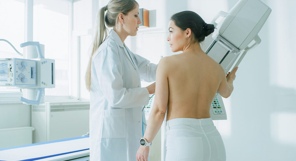
When you have a mammogram, you will need to undress above the waist. A female technologist will position your breasts on the mammogram machine. Your breast is briefly compressed or squeezed between 2 plates attached to the mammogram machine. The technologist must compress your breast to keep it from moving, and to make the layer of breast tissue thinner so it’s easier for the doctor to look through your breast tissue. Although the compression can feel uncomfortable and even painful for some women, it only lasts a few seconds and is needed to get a good picture. Talk to the technologist if you have pain. She can reposition you to make the pressure as comfortable as possible. Although the time you are exposed to x-rays is just seconds, the entire procedure for a mammogram from start to finish takes only about 20 minutes.
All of our mammograms done at Conemaugh are performed on Full-field digital mammography units that capture the picture in a digital format which can be looked at on a computer screen. The benefit of digital mammograms are that the original pictures can be magnified and looked at in many different ways on the computer screen. In addition, several studies have found that digital mammography is more accurate in finding cancers in women younger than 50 and in women with dense breast tissue.
It’s best to find a facility that you are comfortable with and plan to get your regular mammograms there each year. That way, your mammogram pictures are all in one place. If you do have to change facilities, call ahead to find out what you will need to do in order to get your old pictures sent to the new place.
Schedule your mammogram when your breasts are not tender or swollen to help reduce discomfort and get a good picture. If you are still menstruating, try to avoid the week just before your period. Always describe any breast changes or problems you are having to the technologist doing the mammogram. Also, describe any medical history that could affect your breast cancer risk, such as a family history of breast cancer or having prior chest wall radiation therapy. Discuss any new findings or problems in your breasts with your doctor or nurse before having the mammogram.
Being called back for more testing does not mean that you have cancer. In fact, less than 10% of those women called back for more tests are actually found to have breast cancer. Being called back happens fairly often, about 1 in every 10 screening mammograms. It usually just means more pictures or an ultrasound needs to be done to look at a suspicious area more carefully.
The American Cancer Society, the U.S. Department of Health and Human Services (HHS), the American Medical Association (AMA), the National Comprehensive Cancer Network, the National Cancer Institute, the American College of Radiology, the Society of Breast Imagers and many other professional societies recommend annual screening mammograms for every women over the age of 40. Some women schedule the next year’s mammogram and ask to be reminded of the appointment a few weeks ahead of time. Getting a high-quality screening mammogram and having a clinical breast exam on a regular basis are the most effective ways to detect breast cancer early.
Checking one’s own breasts for lumps or other unusual changes is called a breast self-exam, or BSE. This type of exam cannot and should not replace regular screening mammograms or clinical breast exams. Talk to your doctor if you think you may be at higher risk of getting breast cancer to find out what is the best screening test for you.
The American College of Radiology (ACR) has established a uniform way for radiologists to describe mammogram findings. This system, called BI-RADS, includes seven standardized categories, or levels. Each BI-RADS category has a follow-up plan associated with it to help radiologists and other physicians appropriately manage a patient’s care.
For help understanding your mammogram report and your radiologist’s estimate of your cancer risk, visit the American Cancer Society “Understanding your mammogram report – BI-RADS categories.”
If a biopsy is needed, you should discuss the different types of biopsy with your doctor to decide which type is best for you.
Your mammogram report will contain an assessment of breast density. Breast density is based on how much of your breast is made up fatty tissue versus how much is made up of fibrous and glandular tissue. Having dense breasts is normal, but studies have shown that having dense breast tissue will increase your risk of getting breast cancer. We also know that dense breast tissue can make it harder to find cancers on a mammogram. If your breast tissue is heterogeneously dense, there are more areas of fibrous and glandular tissue that can make it hard to see small masses on mammograms. If your breast tissue is extremely dense, this lowers the sensitivity of mammography to detect breast cancers can lead to missing some cancers.
In Pennsylvania and several other states, the women whose mammograms show dense or very breast tissue must be told that they have “dense breasts” in the mammogram report that is sent to these patients. The language used is mandated by state law. For more information regarding your breast density, see the ‘Are you Dense?’ website.
Computer-aided detection (CAD) was developed to help radiologists find abnormal areas on a mammogram by acting as a second set of eyes. Markers appear on the mammogram images which point out areas the radiologist should check more closely. Current research suggests that CAD is not a substitute for experience and expertise in reading mammograms. On going studies are being done to continue to learn about the effects and usefulness of CAD in reading mammograms. All mammograms read at Conemaugh are interpreted with the benefit of CAD.
For most women with private insurance, the cost of screening mammograms is 100% covered without copayments or deductibles, but women should contact their mammography facility or health insurance company for confirmation of the cost and coverage.
Medicare pays for annual screening mammograms for all female Medicare beneficiaries who are age 40 or older. Medicare will also pay for one baseline mammogram for female beneficiaries between the ages of 35 and 39. There is no deductible requirement for this benefit. Information about this coverage is available on the Medicare website or through the Medicare Hotline at 1–800–MEDICARE (1–800–633–4227). For the hearing impaired, the telephone number is 1–877–486–2048.
Information about free or low-cost mammography screening programs is available from the from National Cancer Institute (NCI) at: http://www.cancer.gov/cancertopics/factsheet/detection/mammograms. The NCI’s Cancer Information Service is also available by calling 1–800–4–CANCER (1–800–422–6237). Additional help is available from local hospitals, health departments, women’s centers, or other community groups.
In the United States, mammography is highly regulated. In 1992, Congress passed the Mammography Quality Standards Act (MQSA) to ensure that radiology facilities offering mammography are required to meet minimum quality standards. Today, the US Food and Drug Administration (FDA) certifies every facility offering mammography (except those of the Department of Veterans Affairs). The x-rays are reviewed for quality and information on radiation dose, which is required to be very low.
If the facility meets all of the required standards, the FDA gives its certification. These standards are outlined in the MQSA, which has been in effect since 1994. It is unlawful to do mammograms in the United States without an FDA certificate.
The FDA has a list of all of its certified mammography facilities by state and zip code. You can find those near you by visiting the FDA’s website.
Strict guidelines ensure that mammography equipment is safe and uses the lowest dose of radiation possible. Many people are concerned about the exposure to x-rays, but the level of radiation from a mammogram today does not significantly increase the breast cancer risk for a woman who gets regular mammograms. The dose of radiation that she gets during one screening mammogram is about the same amount of radiation she would average from her natural surroundings (background radiation) over a couple of months. Many studies prove the benefits of mammography and early cancer detection far outweigh any possible harm from the radiation exposure. Women should always let their health care providers and x-ray technologists know if there is any chance that they are pregnant, because radiation can harm a growing fetus.
Although breast cancer screening with yearly mammograms is the best screening test we have now to find cancer early, finding cancer early does not always reduce a woman’s chance of dying from breast cancer. A fast-growing or aggressive cancer may have already spread before it’s found on mammograms. Some mammograms appear totally normal, even though breast cancer is present. Overall, screening mammograms cannot detect about 1 in 5 breast cancers. The main cause of mammograms not being able to show cancer is high breast density. If you feel a lump in your breast, talk to your doctor right away, even if your mammogram is normal. When this happens, you should have another test such as an ultrasound or MRI to make sure there is not an occult cancer (a cancer that cannot be seen on mammograms) in that area. Rarely, palpable lumps must even be biopsied if all the imaging tests are normal, just to be sure that you don’t have breast cancer.
Although breast cancer screening with yearly mammograms is the best screening test we have now to find cancer early, finding cancer early does not always reduce a woman’s chance of dying from breast cancer. A fast growing or aggressive cancer may have already spread before it’s found on mammograms. Some mammograms appear totally normal, even though breast cancer is present. Overall, screening mammograms cannot detect about 1 in 5 breast cancers. The main cause of mammograms not being able to show cancer is high breast density. If you feel a lump in your breast, talk to your doctor right away, even if your mammogram is normal. When this happens, you should have another test such as an ultrasound or MRI to make sure there is not an occult cancer (a cancer that cannot be seen on mammograms) in that area. Rarely, palpable lumps must even be biopsied if all the imaging tests are normal, just to be sure that you don’t have breast cancer.
Screening mammograms are not recommended for average-risk women under the age 40. In some younger women who are at high risk for developing breast cancer (due to a BRCA gene mutation, a strong family history, or other factors), both yearly breast MRIs and mammograms are recommended. For most of these women, screening should begin at 30 years of age. This decision should be based on discussions between patients and their health care providers, taking into account personal circumstances and preferences. This American Cancer Society link Breast Cancer: Early Detection gives more details about the breast cancer screening recommendations. It also tells you more about figuring out your individual breast cancer risk.
For women who have had a mastectomy, mammograms are recommended on the unaffected breast each year. This is very important, because women who have had breast cancer are at higher risk of developing a new cancer in the other breast. For women who have been treated with breast conservation therapy or lumpectomy, yearly mammograms are recommended for both breasts.
Women who have implants are a special challenge for mammogram screening. The x-rays used for imaging the breasts cannot go through silicone or saline implants well enough to show the breast tissue that is over or under it. This means that the part of the breast tissue covered up by the implant will not be seen on the mammogram. In order to see as much breast tissue as possible, women with implants have 4 extra pictures called implant displacement (ID) views, when the implant is pushed back against the chest wall and the breast tissue is pulled forward over it. They are easier in women whose implants are placed underneath (or behind) the chest muscle. Although these women do have more pictures taken at each mammogram, the guidelines for how often women with implants should have screening mammograms are the same as for women without them. Very rarely, mammograms can cause an implant to rupture. It’s very important to tell the technologist before you have your mammogram if you have implants.